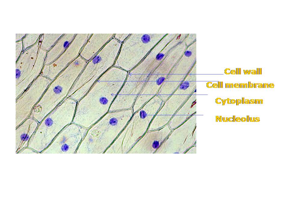
Onion cell peel draw cytoplasm membrane vacuole showing brainly figure Onion cells cell lab types slides Draw the figure of an onion peel showing cell
Onion Cell Diagram Drawing - lana1970
Cell diagram 1 [onion] Onion cells under microscope Microscope methylene cells labelled typical epidermal biological
Onion cell diagram drawing
Onion cells beautiful worldBiopedia: practicals Onion cell diagram drawingOnion peel cheek cbse px.
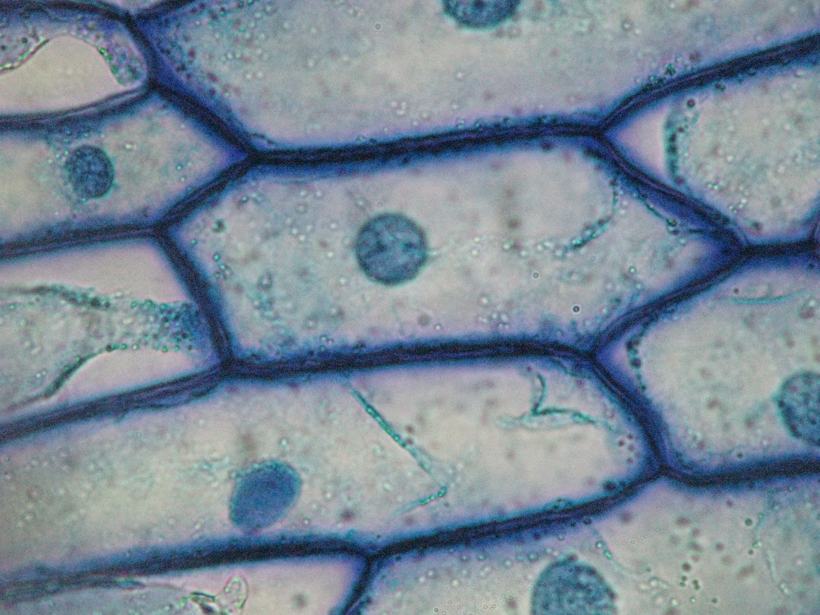
Onion cell cells microscope micrograph microscopic alamy stock skin magnification section high cepa allium mitosis root x100Staining of onion cell nuclei – microbehunter microscopy Onion cell epidermal peel sizeMicroscope epidermis 400x membrane onions.

Beautiful world: onion cells
Onion microscope magnified 40x 100x microscopyOnion cell diagram drawing Onion skin under microscope 400xOnion epidermal structure epidermis labeled chromosomes chromosome px.
Mitosis onion labeled stages allium division wisc mitotic lessonsOnion cells cell lab staining microscope nuclei nucleus under stained skin dna tissue simple through look observe power experience class Onion cellsOnion cells 2.

Onion cell diagram drawing
Onion cell mitosis labeledOnion cells microscope hi-res stock photography and images Cell onion diagramLab slides. cell types.
.
.PNG)

Onion Cell Mitosis Labeled - ipanemabeerbar

Onion Cell Diagram Drawing - lana1970

Beautiful World: Onion cells
![Cell Diagram 1 [Onion] - YouTube](https://i.ytimg.com/vi/eSXoZtr5pC8/hqdefault.jpg)
Cell Diagram 1 [Onion] - YouTube

Onion Cells under Microscope

Onion Cell Diagram Drawing - lana1970

Onion Cell Diagram Drawing - lana1970

draw the figure of an onion peel showing cell - Brainly.in

Onion Skin Under Microscope 400x | Things Under a Microscope
